
Adrenal Gland
| Date of Presentation | 2/18/2025 |
| Attending pathologist | Anil Parwani, MD, PhD, MBA |
| Presented by | Efrain Lee Diaz, MD |
| Organ | Adrenal gland |
HISTORY:
A female patient in her 60’s with known history of right breast fibroadenoma, left breast high grade ductal carcinoma in situ and uterine leiomyosarcoma presented with an adrenal mass.
CT scan that found a new solid heterogenous mass in the suprarenal location (likely an adrenal mass measuring 10.5 x 5.8 x 6.9 cm) that caused mass effect on the left kidney, and displacing the left renal vein.
The patient was admitted for a retroperitoneal mass resection en block with left adrenalectomy.
GROSS:
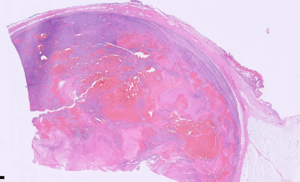
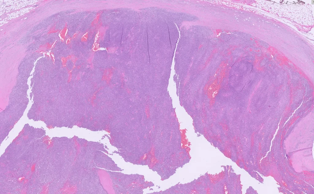
Cut section through the mass reveals dark red to yellow-tan, diffusely necrotic and hemorrhagic tissue that is partially lobulated. The mass is approximately 90% necrotic.
Rim of possible adrenal gland tissue that is 1.8 x 0.9 x 0.2 cm that abuts the mass.
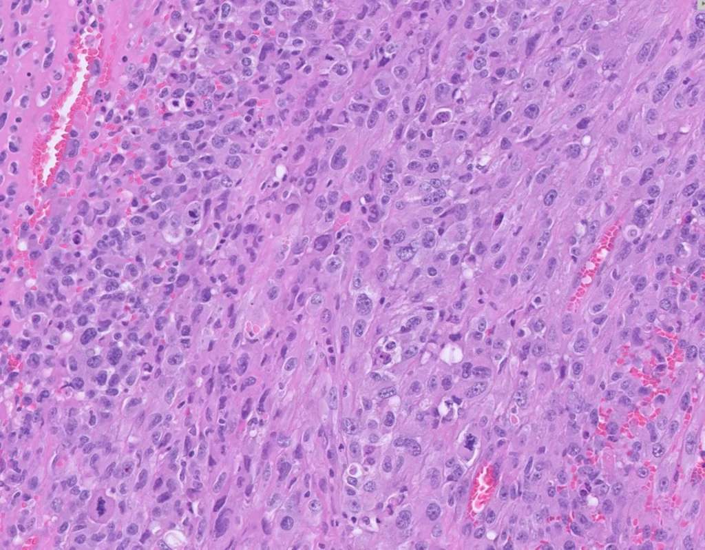

What is the most likely diagnosis?
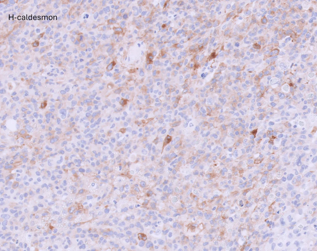
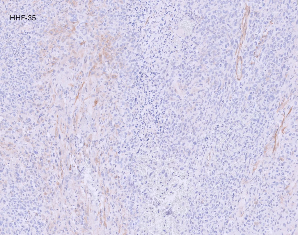
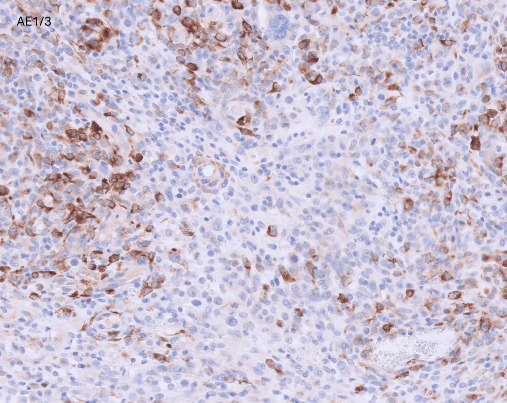
- Metastatic uterine leiomyosarcoma involving the adrenal gland
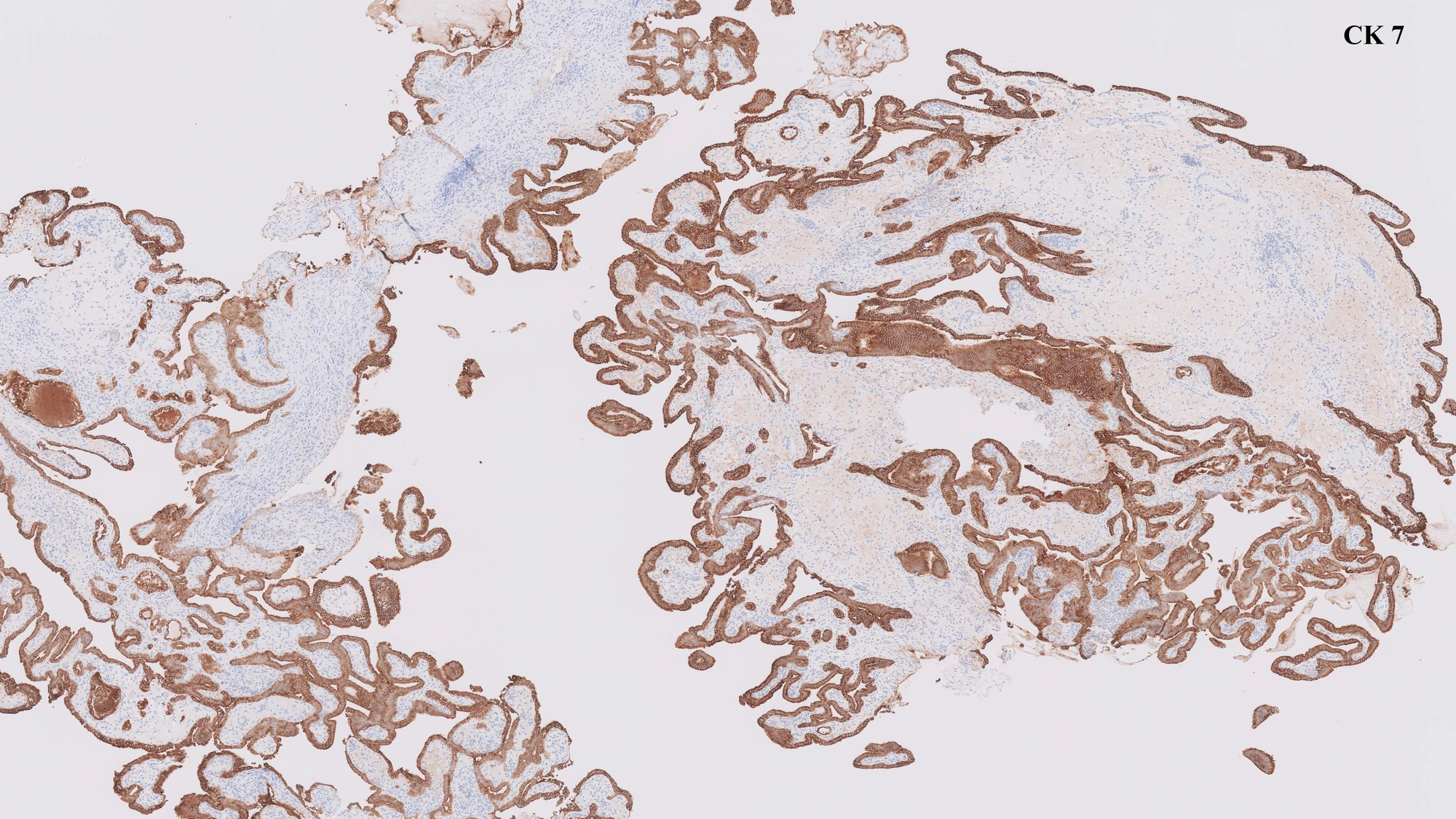
- Metastatic breast carcinoma involving the adrenal gland
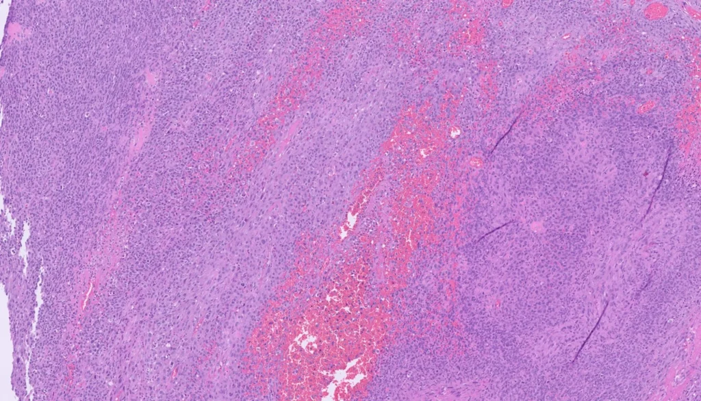
- Primary sarcoma arising in the adrenal
- Sarcomatoid carcinoma of renal origin